Mole screening, dermatoscopy, or dermoscopy is a non-invasive diagnostic method for observing the skin’s surface, including all moles and other existing pigmentation. The dermatologist conducts the screening using a specialized microscopic technique with an instrument called a dermoscope.
Dermoscopy, dermatoscopy, or melanomascopy are different professional terms that refer to the same procedure – mole screening by a dermatologist. Whether it’s a routine annual check-up, a mole injury, or a change in its appearance, dermoscopy will alleviate any doubts.
What is a mole dermoscopy?
The dermoscope magnifies the skin’s surface several tens of times using polarized or non-polarized light. This allows for the observation and evaluation of all surface and deeper layer changes in the upper dermis. Consequently, all changes can be observed.
Dermoscopy is highly beneficial in detecting imperceptible changes within moles that occur deeper in the skin. This enables the determination of whether a particular mole is at risk. Additionally, this screening is crucial for early melanoma diagnosis.
As a diagnostic procedure in dermatology, mole screening dates back to 1664 when the initial microscopic screening of the skin were documented. Subsequently, the German dermatologist Johann Safier introduced the use of light in skin change observation, while the dermatologist Goldman coined the term “dermatoscopy.” Following the First World Congress of Dermatoscopy in 2001, the official professional term for mole examination became dermoscopy.
How to tell the difference among moles?
Moles, also known as nevi, are benign growths that can occur on any part of the body. They can be either congenital or acquired throughout life due to various factors. Benign moles consist of melanin, the dark pigment cells responsible for the skin’s color. However, melanoma cells, which are skin tumors, have altered DNA and multiply abnormally throughout the skin, including in benign moles.
In women, existing moles can undergo changes and numerous new moles can appear during pregnancy, influenced by melanocyte-stimulating hormone (MSH), estrogen, and progesterone.
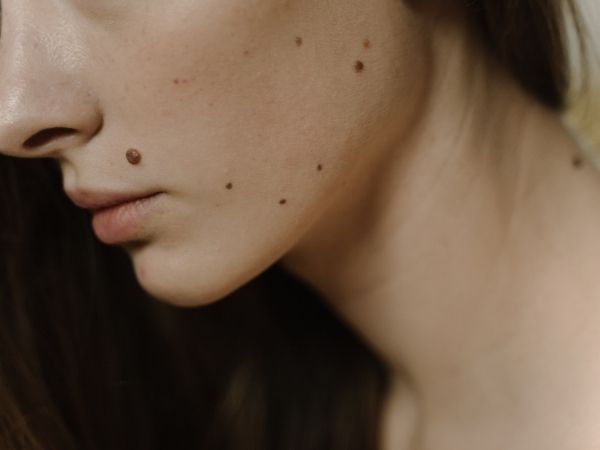
The color of moles can range from pink to light and dark brown. They can be flush with the skin, flat or raised, pedunculated, rough, smooth, soft, or hard. Their diameter typically ranges from several millimeters to several centimeters. On average, adults have between 10 and 40 moles on their bodies by the age of thirty. After the age of fifty, new moles rarely develop.
Benign moles
The majority of moles on the body are benign, meaning they are harmless. So, how can we identify them? Here are the visible characteristics:
- Harmless moles are typically symmetrical in shape, appearing oval, round, or oblong.
- Their color ranges from light brown to dark brown.
- The edges of these moles are straight and well-defined.
- The diameter of a mole is usually not larger than 6 mm, although size alone is not a cause for concern.
However, if any of the following changes occur, it may indicate a potential risk, and it is important to seek advice from a dermatologist.
Risky moles
Although moles are typically benign skin growths, there are instances where they can undergo risky changes that are not visible to the naked eye. In some cases, these moles may also contain cancerous cells. When you notice any of the following changes, it is crucial to seek medical advice:
- The mole has an unusual, irregular shape, with jagged or uneven edges and varying colors.
- Pay attention to any sudden changes in the mole’s color, particularly if it becomes very dark, almost black. Consulting a dermatologist is recommended in such cases.
- If the mole becomes raised above the skin surface, instead of being flush with it.
- Any signs of wetness, bleeding, inflammation, or other visible alterations require a visit to a dermatologist.
- Moles with a diameter greater than 6 mm, along with other noticeable changes.
Unfortunately, the incidence of skin cancer is increasing every year, surpassing all other types of cancers combined.
Melanoma, one of the rarest but most aggressive forms of skin cancer, primarily affects individuals aged 25 to 29 years. It typically develops from moles or atypical moles and rarely arises directly from the skin. Harmful UV radiation, especially in individuals with a history of sunburn, is a significant contributing factor to these cell mutations.
Atypical moles can closely resemble melanoma and exhibit characteristics such as asymmetry, changes in pigmentation, and black or gray coloration. In such cases, a biopsy is performed to rule out the possibility of melanoma.
If you have noticed any of the aforementioned changes in your existing moles, it is advisable to visit a dermatologist. Through a screening, they can determine whether you have so-called atypical or dysplastic moles.

Why is a mole dermoscopy important?
We would like to emphasize the utmost importance of making annual mole screening a regular habit. Just like regular dental check-ups or annual check-ups with a gynecologist, this type of examination should be prioritized and not skipped or postponed. It is evident that any peculiar or abnormal changes in the color or shape of a mole, the sudden appearance of numerous new moles, bleeding, or mole injuries necessitate a visit to a dermatology specialist.
All the advantages of dermoscopy
In addition to being quick and simple, dermoscopy provides valuable assistance to dermatologists in the following ways:
- It enables faster and easier monitoring of pigment changes on the skin.
- It helps in accurately determining the nature of skin changes, distinguishing between pigmentary changes and melanoma, and identifying changes that require removal. Previously, any suspicious skin change required surgical intervention and histopathological analysis, but with the use of dermoscopy, the need for such interventions has significantly reduced.
- Dermoscopy is highly beneficial for distinguishing between melanocytic and non-melanocytic cells, which is crucial for early melanoma diagnosis. Early detection and treatment of melanoma can lead to successful outcomes.
- Dermoscopy aids in distinguishing different types of skin tumors, facilitating faster and easier diagnoses.
- It allows for the monitoring and analysis of suspicious moles over time and facilitates their removal when necessary.
- Regular dermoscopy is particularly important for individuals at a higher risk of developing melanoma, such as those with a personal history of melanoma or other skin tumors, or those with a family history of the disease.
- Dermoscopy is also beneficial for individuals with a large number of moles (more than 50), those who are highly sensitive to sunlight, individuals who frequent tanning beds, or those who have had sunburns during childhood or adolescence.
- Dermoscopy is also employed for warts screening and diagnosing fungal skin infections.
When should a dermoscopy be performed?
Dermoscopy is typically performed alongside a clinical dermatological examination, especially when the screening alone is insufficient for an accurate diagnosis. The dermatologist may recommend additional diagnostic methods such as skin biopsy and mole screening.
To ensure overall skin health and minimize the risk of melanoma or other skin changes, it is recommended to undergo preventive mole screening once a year. The following groups are especially encouraged to have regular mole dermoscopy:
- Pregnant women, as they may experience various degenerative skin conditions and an increased number of moles during the second trimester.
- Children, during periods of accelerated growth when pigment changes may occur on the skin.
- Individuals at higher risk, including those with more than 50 moles or those with atypical moles.
- People who consciously expose themselves to harmful UV radiation, such as those who visit tanning salons or spend extended periods in the sun due to occupational reasons.
- Individuals with a family history of melanoma or other skin cancers, or those with certain predisposing skin conditions, should undergo annual mole screening.
- Mole dermoscopy is mandatory if there have been any changes in the color, shape, or number of moles, as well as if there has been an injury to a mole, itching, pain, redness, bleeding, or moisture in the affected area.
What does a mole dermoscopy look like?
Mole dermoscopy is a simple and non-invasive diagnostic method that allows for precise identification and characterization of pigment changes on the skin. It involves the use of a handheld instrument called a dermoscope, which magnifies the observed area 10 to 14 times.
Dermoscopy provides visual enlargement and illumination of the skin surface, either in its entirety or specific areas. Prior to the screening, the dermatologist applies oil to the skin and then places the dermoscope on the desired area to observe the magnified surface.
With digital dermoscopy, various skin changes can be captured through photography. These photos are stored and used during follow-up examinations to monitor any potential changes on the skin.
Dermoscopy is a completely safe, so there are no risks or the possibility of any complications. This type of skinv screening is completely painless. It lasts from a few minutes to half an hour, depending on the surface of the skin that the doctor needs to examine.
Before mole dermoscopy
To prepare for the screening, it is advisable to perform a self-examination of your skin and moles at home. This way, you can identify any changes you have noticed and bring them to the doctor’s attention, if necessary.
How accurate is dermoscopy?
When discussing melanoma, it can be visually detected without any instruments in up to 65 percent of cases. However, with a dermoscopy, the reliability of assessment increases to up to 95 percent. That is why preventive mole dermoscopy is crucial. Detecting melanoma at an early stage greatly enhances the chances of successful treatment. Dermoscopy allows for the observation of changes in the skin’s papillary dermis, which is the layer just beneath the surface epidermis. Changes occurring deeper than that cannot be detected through dermoscopy.
What after mole dermoscopy?
After the mole screening, you can resume your daily activities immediately. Taking into account all the factors and potential risks, the dermatologist will decide whether further measures are necessary and what they entail. If deemed necessary, one of the accompanying procedures may involve the surgical or radio-wave removal of moles. Following the removal, the mole is sent for histopathological examination to determine the possibility of malignancy. Any healthy mole that may cause discomfort or be prone to friction and irritation can be safely removed without risking one’s health.
You might not know this about moles
- When we are born, we have very few moles on our skin, known as congenital naevi.
- The highest number of moles typically appear during puberty and are referred to as acquired naevi. In males, moles mostly develop by the age of 25, while in females it is by the age of 28 or 30.
- The hormonal influence during pregnancy often leads to the symptomatic appearance of a greater number of moles.
- Individuals with a mole above their lip are often considered attractive.
- The famous mole on Marilyn Monroe’s cheek has become her trademark.
Price and additional information
You can see the prices on the Pricelist page. We believe that there is always a question you want to ask our doctors, so know that we are always here for you. Call us at phone number 064/237-0707 or write to us at imedic.rs@gmail.com.